AZD5991 Selective Mcl-1 Inhibition Drives Endoplasmic Reticulum Stress and Immunogenic Cell Death
Copy code to add this image to your page
Targeting the anti-apoptotic B-cell lymphoma-2 (Bcl-2) protein family represents a key therapeutic strategy in oncology. Among these family members, Myeloid cell leukemia 1 (Mcl-1) serves as a primary survival factor, enabling tumor cells to evade programmed cell death. AZD5991 is a potent, small-molecule BH3-mimetic designed to selectively bind the hydrophobic groove of Mcl-1. This binding disrupts interactions with pro-apoptotic BH3-only proteins, including Bim and Noxa. Under physiological and pathological conditions, Mcl-1 overexpression frequently confers resistance to standard chemotherapeutic agents and monotherapy Bcl-2 inhibitors. Consequently, neutralizing Mcl-1 restores apoptotic sensitivity across multiple hematologic and solid tumor models.
In colorectal cancer models, single-agent suppression of anti-apoptotic proteins often proves insufficient due to redundant survival signaling nodes. Concurrent inhibition of Mcl-1 using AZD5991, alongside the dual Bcl-2/Bcl-XL antagonist ABT-263, forces extensive reorganization of apoptotic machinery. When combined with PF-543—a selective inhibitor of sphingosine kinase 1 (SphK1)—this tripartite regimen initiates a distinct pathway leading to immunogenic cell death (ICD). Rather than executing silent, non-inflammatory apoptosis, tumor cells undergoing this combination treatment display biochemical hallmarks of immune engagement.
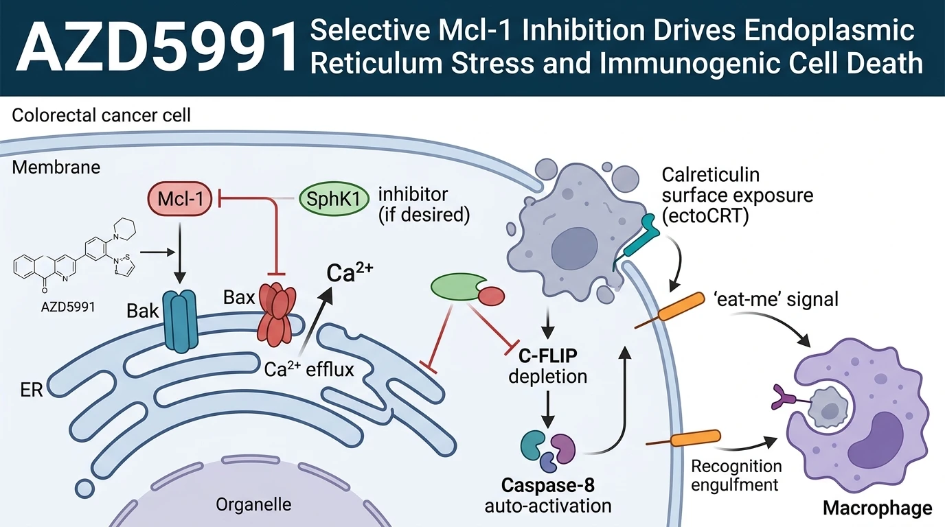
Mechanistically, joint disruption of Mcl-1 and Bcl-2/Bcl-XL mobilizes the multidomain pro-apoptotic effectors Bak and Bax at the endoplasmic reticulum (ER) membrane. Under normal conditions, Mcl-1 sequesters Bak at both mitochondrial and ER membranes. Displacement of Bak by AZD5991 induces localized ER membrane permeabilization, precipitating a rapid release of luminal Ca2+ into the cytosol. Elevated cytosolic calcium levels trigger downstream cellular stress cascades. However, full execution of the immunogenic pathway requires concurrent attenuation of lipid survival signaling.
Sphingosine kinase 1 generates sphingosine-1-phosphate (S1P), a bioactive lipid messenger that reinforces cell survival during metabolic stress. Elevated SphK1 activity stabilizes cellular FLICE-like inhibitory protein (c-FLIP), an endogenous inhibitor of Caspase-8 activation. In colorectal cancer cells treated with PF-543, inhibition of SphK1 suppresses S1P production. This biochemical blockade induces rapid proteasomal degradation of c-FLIP. Depletion of c-FLIP relieves the tonic suppression of Caspase-8, facilitating its auto-catalytic processing and activation at the cytosolic face of the ER.
Activated Caspase-8 acts on structural and chaperone substrates within the cell. Crucially, Caspase-8 activity leads to the structural modification and dimerization of calreticulin, a lumenal ER chaperone protein. Dimerized calreticulin rapidly translocates across the plasma membrane to form cell surface complexes, a process termed ecto-calreticulin exposure (ectoCRT). In vitro assay models demonstrate that ectoCRT serves as an essential eat-me signal for innate immune surveillance. Professional antigen-presenting cells, such as macrophages and dendritic cells, recognize ectoCRT via low-density lipoprotein receptor-related protein 1 (LRP1).
In preclinical murine models, treatment with AZD5991 and targeted co-inhibitors promoted enhanced macrophage-mediated phagocytosis. Immunogenic clearance of apoptotic debris induced durable anti-tumor T-cell responses in vivo. Notably, the induction of ectoCRT and subsequent phagocytic clearance remained dependent on the simultaneous suppression of SphK1 and anti-apoptotic Bcl-2 proteins. Isolated Mcl-1 inhibition without SphK1 blockade failed to induce equivalent levels of surface calreticulin exposure. These findings define a specific pharmacological axis wherein targeting Mcl-1 and SphK1 converges on ER stress pathways to drive productive immunogenic clearance.
Pharmacological Synergism and Immunogenic Cell Death Execution
Copy code to add this image to your page
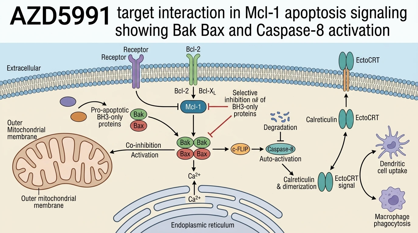
AZD5991 acts as a selective BH3-mimetic inhibitor of Mcl-1. When combined with ABT-263 and PF-543, it triggers Bak/Bax oligomerization at the endoplasmic reticulum. SphK1 inhibition via PF-543 blocks S1P synthesis, causing c-FLIP degradation. Unrestrained c-FLIP depletion promotes Caspase-8 cleavage and subsequent calreticulin dimerization. Surface exposure of calreticulin (ectoCRT) signals phagocytic clearance by macrophages, effectively transforming apoptotic stress into an immunogenic tumor response.
Endoplasmic Reticulum Calcium Mobilization and Caspase Activation
Dual disruption of Mcl-1 and Bcl-2/Bcl-XL via AZD5991 and ABT-263 alters organelle ion homeostasis. Under tested in vitro assay conditions, Bak and Bax oligomerize at the endoplasmic reticulum membrane. Ca2+ ions efflux rapidly into the cytoplasm within hours of target engagement. This sudden ion flux triggers cellular stress responses dependent on lipid signaling status. In control conditions, sphingosine kinase 1 activity preserves c-FLIP levels to prevent ectopic Caspase-8 processing. Pharmacological blockade with PF-543 degrades c-FLIP efficiently. Caspase-8 undergoes auto-proteolytic cleavage directly following c-FLIP clearance; this enzymatic cascade operates independently of canonical death receptor engagement.
Calreticulin Translocation Enhances Macrophage Phagocytosis in Vivo
Surface expression of calreticulin represents a critical biochemical prerequisite for immunogenic cell death. Cleaved Caspase-8 facilitates the dimerization and rapid externalization of calreticulin to the outer lipid bilayer. In patient-derived colorectal cell line models, ectoCRT exposure reached peak levels following co-treatment with AZD5991, ABT-263, and PF-543. Phagocytosis assays confirmed accelerated engulfment by bone marrow-derived macrophages. Within immunocompetent murine xenograft models, this triple combination reduced primary tumor burden relative to monotherapy cohorts. Preliminary data suggest that the resulting immune response relies on cytotoxic T-cell recruitment following macrophage activation. Therapeutic efficacy in human clinical settings remains to be confirmed.