L-Kynurenine AhR agonist signaling regulates IDO1 degradation through CUL4BAhR ubiquitin ligase
Copy code to add this image to your page
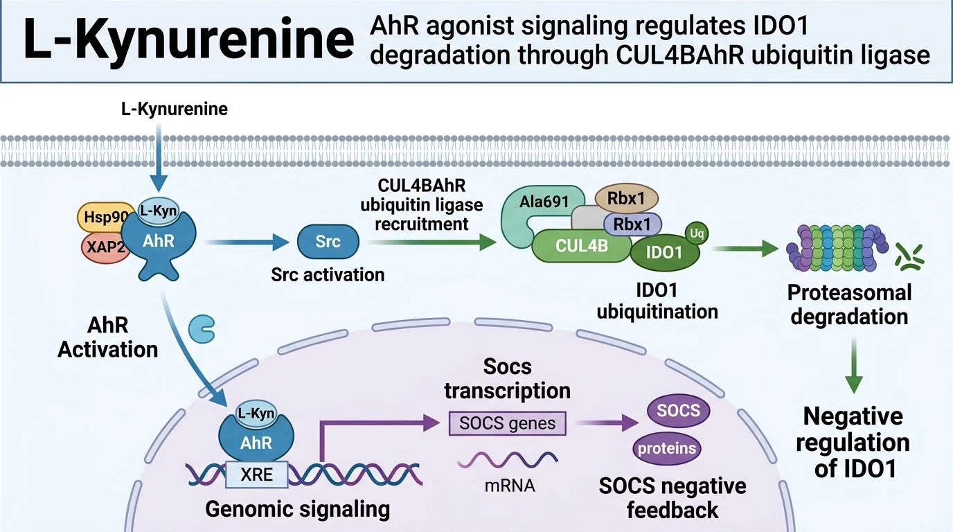
Indoleamine 2,3-dioxygenase 1 (IDO1) catalyzes the rate-limiting initial step in the kynurenine pathway, degrading the essential amino acid L-tryptophan into N-formylkynurenine, which rapidly converts to L-Kynurenine. Downstream accumulation of L-Kynurenine alters local metabolic microenvironments, suppressing effector T-cell responses while favoring regulatory T-cell differentiation. Operating beyond a passive metabolic byproduct, L-Kynurenine actively functions as an endogenous ligand for the aryl hydrocarbon receptor (AhR), a basic helix-loop-helix transcription factor retained in the cytoplasm by a chaperone complex containing HSP90, XAP2, and p23.
Upon intracellular concentration thresholds being reached, L-Kynurenine occupies the PAS-B ligand-binding domain of AhR. As an endogenous L-Kynurenine AhR agonist, it induces a conformational rearrangement that exposes the receptor's nuclear localization signal. This structural shift promotes chaperone dissociation and facilitates transport across the nuclear pore complex. Inside the nucleus, AhR heterodimerizes with the AhR nuclear translocator (ARNT). The resulting complex binds specific canonical dioxin response elements (DREs) or xenobiotic response elements (XREs) across genomic regulatory regions. This engagement recruits transcriptional coactivators to initiate transcription of target genes, including Ido1, Socs2, and Socs3.
Simultaneously, ligand engagement activates a non-genomic cascade that operates independently of classical DRE-driven gene transcription. Receptor activation prompts the release and catalytic activation of c-Src tyrosine kinase from the cytosolic chaperone complex. Active c-Src targets specific immunoreceptor tyrosine-based inhibitory motifs (ITIMs) located on the intracellular polypeptide chain of IDO1. Phosphorylation of ITIM residues, specifically Tyr115 and Tyr158, converts IDO1 into a functional signaling scaffold capable of recruiting SH2 domain-containing proteins.
Concurrently, non-genomic activation converts cytosolic AhR into a structural subunit of a Cullin-RING E3 ubiquitin ligase apparatus. AhR incorporates into the CUL4B-based E3 ligase machinery (CUL4BAhR), forming an active protein complex comprising CUL4B, RBX1, Elongin B, Elongin C, and target recognition components. The phosphorylated ITIM motifs on IDO1 serve as direct binding surfaces for this newly assembled ligase complex. Once docked, the CUL4BAhR ligase transfers ubiquitin moieties from E2 conjugating enzymes onto lysine residues of IDO1.
Polyubiquitination targets phosphorylated IDO1 to the 26S proteasome for catalytic degradation. Destruction of the enzyme decreases cellular IDO1 protein abundance and halts ongoing tryptophan catabolism. This sequential mechanism operates as an intrinsic negative feedback loop: IDO1 activity generates L-Kynurenine, which subsequently triggers the non-genomic ubiquitin ligase assembly required to degrade IDO1. In murine dendritic cell cultures and human cell lines under physiological conditions, this regulatory loop limits hyper-activation of the kynurenine pathway, preventing prolonged local tryptophan depletion while fine-tuning baseline immunoregulatory phenotypes.
Pharmacological Feedback Dynamics and Non-Genomic Signal Transduction
Copy code to add this image to your page
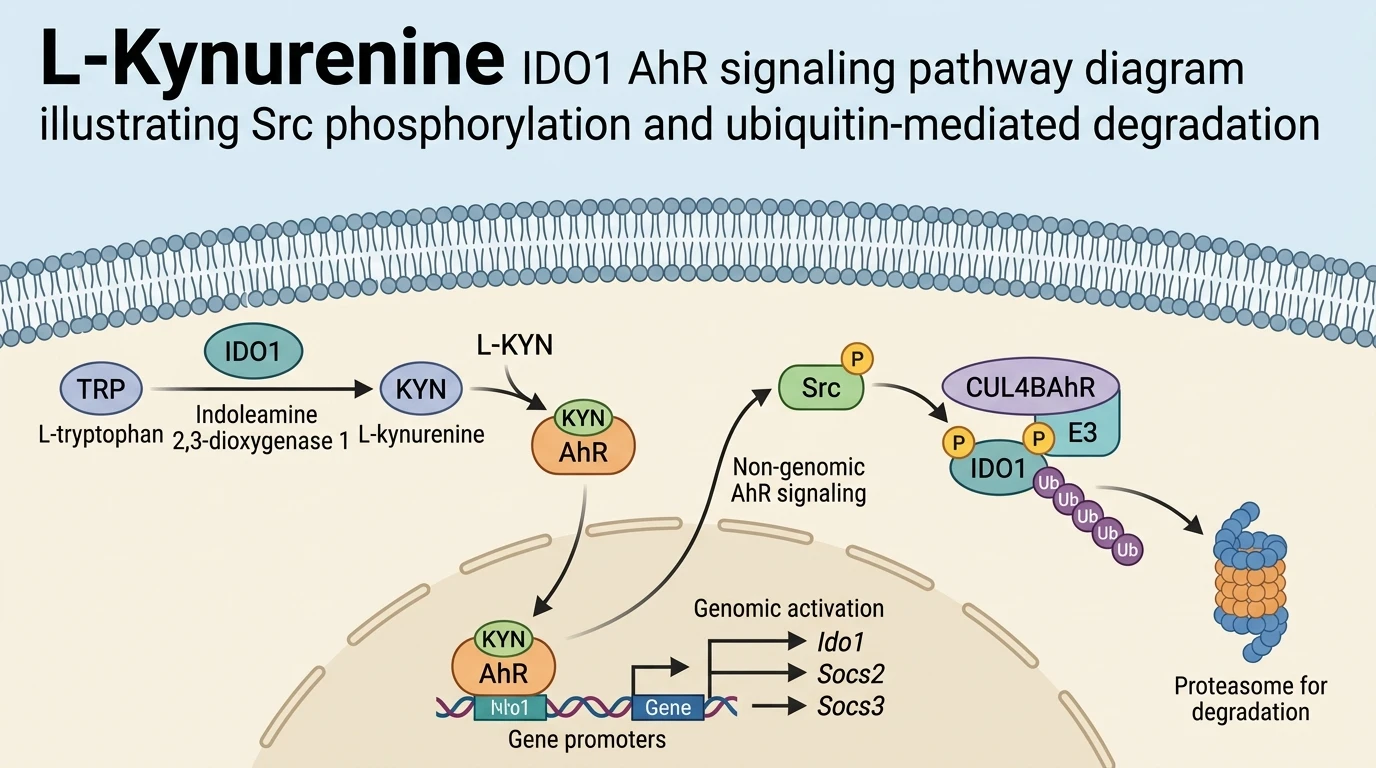
L-Kynurenine functions as an endogenous ligand for the aryl hydrocarbon receptor (AhR), driving both genomic transcription and non-genomic E3 ubiquitin ligase activity. Ligand binding prompts AhR to direct transcription of genes including Ido1, Socs2, and Socs3. Simultaneously, non-genomic AhR signaling recruits Src kinase to phosphorylate IDO1, enabling assembly of the CUL4BAhR E3 ubiquitin ligase complex. This dual mechanism induces polyubiquitination and proteasomal degradation of IDO1. The resulting negative feedback loop quenches enzymatic conversion of tryptophan, modulating dendritic cell immunoregulatory phenotypes in specific cellular environments.
Src Kinase Phosphorylation Triggers IDO1 Ubiquitin Ligase Assembly
Src kinase activation occurs within minutes of ligand engagement. Under tested in vitro assay conditions, binding of the L-Kynurenine AhR agonist destabilizes cytosolic chaperone interactions with the receptor complex. Release of c-Src initiates targeted phosphorylation. Specifically, Tyr115 and Tyr158 motifs on IDO1 undergo rapid phosphorylation. Phospho-specific western blotting confirmed elevated signal intensity within fifteen minutes. These modified residues function as docking platforms for downstream ligase components. Blockade of Src catalytic activity with small-molecule inhibitors completely halted IDO1 modification. Consequently, structural assembly of the ubiquitin ligase machinery requires primary tyrosine phosphorylation.
CUL4BAhR E3 Ligase Complex Drives Proteasomal Turnover
The assembly of CUL4BAhR relies on ligand-induced conformational alterations. Within primary dendritic cell assays, active ligase complexes transfer polyubiquitin chains directly onto IDO1. Lysine 48-linked polyubiquitination marks the enzyme for proteasomal destruction. Co-immunoprecipitation assays verified physical association between CUL4B and AhR post-activation. Cycloheximide chase experiments demonstrated a reduced IDO1 protein half-life following metabolite exposure. Pretreatment with the proteasome inhibitor MG132 fully rescued IDO1 expression levels. Thus, post-translational degradation effectively quenches enzymatic conversion of tryptophan under specific metabolic stress conditions.
Transcriptional Induction of Socs Genes Enforces Immunosuppressive Reset
Genomic signaling proceeds alongside non-genomic ubiquitin ligase activity. Nuclear AhR-ARNT heterodimers bind target XRE sequences within two hours of stimulation. In vitro reporter assays demonstrated robust upregulation of Socs2 and Socs3 transcription. Elevated SOCS protein levels suppress baseline STAT phosphorylation events. This attenuation modulates dendritic cell maturation markers in cell culture models. Flow cytometry revealed reduced surface expression of CD80 and CD86 post-treatment. Observed immunoregulatory shifts remain dependent on cellular contextual factors and local amino acid availability.