PACAP 1-38 and PACAP 138 from Selleck modulate VTAPBP neurons to attenuate trigeminovascular nociception
Copy code to add this image to your page
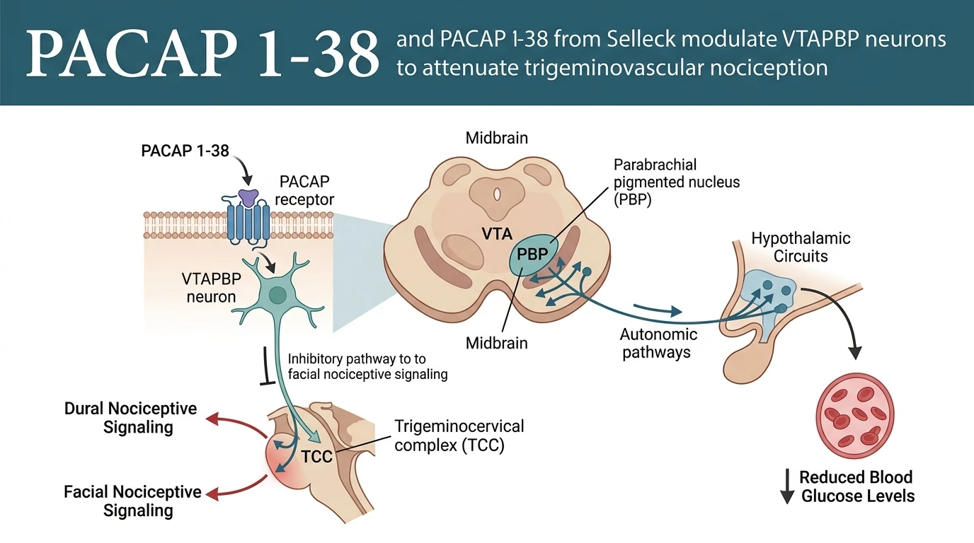
The ventral tegmental area parabrachial pigmented nucleus (VTAPBP) functions as a key midbrain nexus integrating reward processing, autonomic regulation, and trigeminovascular sensory modulation. Pituitary adenylate cyclase-activating polypeptide (PACAP) acts within this structure through three distinct class B G-protein coupled receptors: PAC1, VPAC1, and VPAC2. Microinjection of PACAP38 directly into the VTAPBP produces pronounced antinociceptive effects, significantly dampening both spontaneous activity in trigeminovascular neurons and evoked responses within the trigeminocervical complex (TCC). These inhibitory actions affect TCC neuronal responses triggered by dural electrical stimulation as well as noxious mechanical facial pinching in anesthetized rat models.
Understanding how VTAPBP neurons regulate trigeminovascular processing requires evaluating local pharmacology and receptor engagement. Comparative microinjection studies reveal that PACAP 1-38 administration mirrors the actions of selective monoaminergic agents, including the 5-HT1B/1D agonist naratriptan and the dopamine D2 agonist quinpirole. Local infusion of each ligand into the VTAPBP suppresses dural-evoked electrical discharges and noxious mechanical sensory transmission. This convergence demonstrates that midbrain PACAP receptor activation engages descending inhibitory circuitry rather than facilitating peripheral nociceptive signaling.
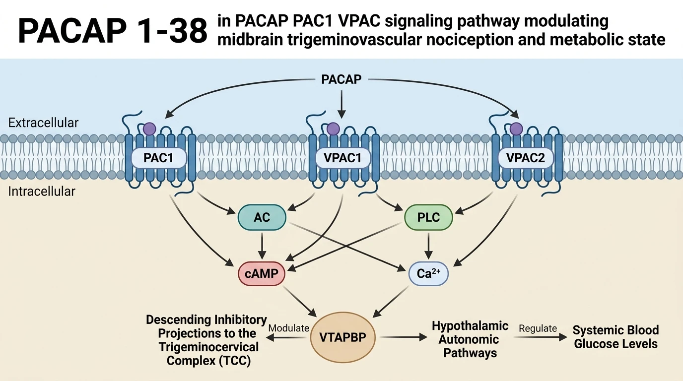
At the cellular level, PAC1 receptor activation stimulates adenylyl cyclase via Gαs coupling, raising intracellular cyclic AMP (cAMP) and activating protein kinase A (PKA). Concurrently, PAC1 activation engages Gαq-mediated phospholipase C (PLC) signaling, driving intracellular calcium release. VPAC1 and VPAC2 receptors similarly elevate cAMP but display equal affinities for PACAP and vasoactive intestinal peptide (VIP). Within VTAPBP microcircuits, these downstream biochemical cascades modify neuronal membrane potential and synaptic transmission. The resulting alterations in output neuron firing modulate descending inhibitory control over primary trigeminal afferents in the TCC.
In vivo electrophysiological studies in Sprague-Dawley rats provided direct measurement of these physiological responses. Extracellular single-unit recordings from second-order TCC neurons demonstrated a marked reduction in background firing frequency following VTAPBP microinjections. Baseline spontaneous firing decreased within fifteen minutes post-injection. Dural electrical stimulation (0.5 Hz, 2–5 mA, 1 ms pulse duration) elicited baseline action potential trains that were significantly attenuated after PACAP38 application. Responses to noxious mechanical pinch applied to the ophthalmic (V1) receptive field decreased in parallel.
Parallel to its sensory actions, pharmacological modulation of VTAPBP neurons alters systemic metabolic parameters. Microinjection of PACAP38 into the VTAPBP causes a measurable reduction in circulating blood glucose levels. This metabolic shift points to functional crosstalk between midbrain nociceptive integration and central glucose regulation. The VTAPBP sends anatomical projections to hypothalamic structures involved in energy balance, such as the ventromedial hypothalamus and lateral hypothalamic area. PACAP receptor engagement within the VTAPBP likely modulates descending autonomic tone, influencing peripheral glucose utilization or hepatic glucose production.
In experimental protocols, consistent target engagement depends on ligand stability and purity. Standardized peptides like PACAP 138 from Selleck enable precise functional evaluation of PAC1, VPAC1, and VPAC2 signaling pathways across electrophysiological and metabolic assays. The observed dual effects—suppression of TCC nociceptive firing and reduction of blood glucose—indicate that VTAPBP neurons coordinate head pain pathways with metabolic homeostasis. These empirical findings establish midbrain PACAP receptor signaling as an important site of convergence for sensory filtering and neuroendocrine regulation.
VTAPBP Receptor Modulation of Trigeminovascular Nociception and Metabolism
Copy code to add this image to your page
Microinjection of PACAP38 into the ventral tegmental area parabrachial pigmented nucleus (VTAPBP) activates PAC1, VPAC1, and VPAC2 receptors, triggering descending inhibitory control over the trigeminocervical complex (TCC). This activation significantly decreases baseline spontaneous activity in trigeminovascular neurons and inhibits TCC firing evoked by dural electrical and facial mechanical noxious stimuli. Concurrently, VTAPBP activation modulates autonomic signaling to reduce circulating blood glucose levels. These findings demonstrate that midbrain PACAP receptor signaling coordinates central trigeminovascular sensory processing with systemic metabolic homeostasis.
VTAPBP Microinjection of PACAP38 Suppresses Trigeminocervical Neuronal Firing
Local administration of PACAP 1-38 into the VTAPBP produced immediate electrophysiological changes in the trigeminocervical complex. Baseline spontaneous activity in TCC neurons decreased markedly. Dural-evoked electrical responses dropped following peptide infusion. Noxious mechanical stimulation of facial receptive fields similarly showed attenuated action potential output. These observations suggest that midbrain PACAP receptor activation recruits descending inhibitory signaling. Comparable suppression occurred with local microinjection of naratriptan or quinpirole. Downstream trigeminovascular responses remained suppressed throughout the observation window. Applying reliable peptide formulations such as PACAP 138 from Selleck ensured consistent microinjection dosing across experimental rat cohorts. The observed inhibition confirms that VTAPBP signaling modulates central sensory gating.
Central PACAP Signaling in VTAPBP Reduces Systemic Blood Glucose Levels
Pharmacological manipulation of VTAPBP neurons altered systemic metabolic indicators alongside nociceptive parameters. Microinjection of PACAP38 caused a measurable decline in circulating blood glucose levels. Baseline glycemic metrics shifted significantly within thirty minutes post-infusion. This metabolic response highlights a direct link between midbrain pain circuits and neuroendocrine homeostasis. Receptor activation across PAC1, VPAC1, and VPAC2 nodes in the VTAPBP likely modulates autonomic projections extending to hypothalamic metabolic centers. Utilizing validated compounds like PACAP 138 from Selleck allowed accurate characterization of these metabolic dynamics without non-specific vehicle confounders. Systemic glucose alterations remained stable under tested anesthetic conditions. These findings demonstrate that midbrain trigeminovascular control intersects with central regulation of blood glucose.